Coronary Computerised Tomographic Angiogram (Coronary CTA)
CAC imaging is usually complemented by CT coronary angiography (coronary CTA). This is a non-invasive, relatively painless radiological heart imaging of the coronary arteries (i.e. blood vessels that supply oxygenated blood to the heart muscles) for screening, risk stratification and detection of blockages in the coronary arteries.
Coronary CTA can :
- Detect cholesterol build-up (atheromatous plaques) within the coronary arteries
- Assess the degree of narrowing of coronary arteries both within the vessel lumen and within the vessel wall ( i.e. vessel expansion and remodelling)
- Look for coronary artery congenital abnormalities e.g abnormal origin and course of the coronary arteries which can predict sudden death, abnormal communication between coronary artery and the veins, dilatation or swelling of coronary arteries as a results of weakening of the walls of the vessel by cholesterol deposition (termed ectasia)
When a coronary CTA is ordered, it is usually done in 2 steps :
Step 1 :
Perform coronary artery calcium imaging and scoring and assessing the CAC score
Step 2 :
Using intravenous infection of a contrast dye through a small plastic needle into the vein of the arm and performing a CT scan of the heart and its coronary arteries.
The CT scan captures high resolution images of the lumen of the coronary arteries as well as the presence of calcified and/or “soft plaque” in the walls of the arteries.
Present computed tomography coronary angiography images are acquired using very low tube voltage and current and the effective radiation dose can be as low as 0.3 mSv (equivalent of only 10 chest x-rays). The image quality is very good with 97% interpretable segments and a high diagnostic accuracy.
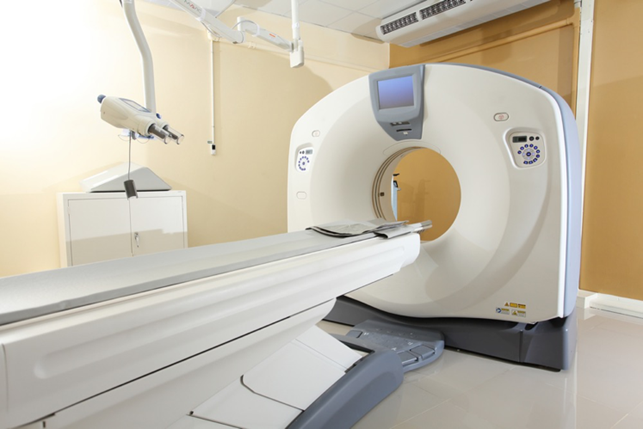
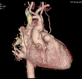
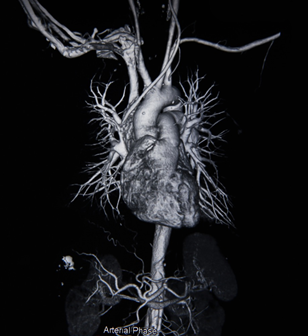
Figure 1 and 2. Coronary CTA showing 3-D rendering of he heart, aorta and coronary arteries
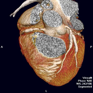
Figure 3. Coronary CTA showing 3-D rendering of the coronary arteries with 3 stents deployed
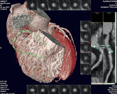
Figure 4. Coronary CTA showing 3-D rendering of the coronary arteries with contrast-like images in longitudinal and cross-section views
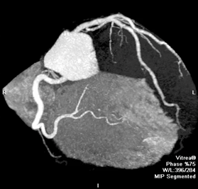
Figure 6. Coronary CTA showing the coronary arteries with contrast-like images.
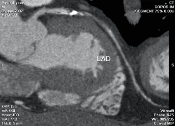
Figure 5. Coronary CTA showing the left front artery with a mixed soft and calcified cholesterol plaque causing significant narrowing of the artery.